6th March 2009
This page contains unofficial supplementary information for the paper:
The following high-resolution versions of the figures from the sauropod neck-posture paper are for the benefit of scientists and reporters. Feel free to reproduce or modify these for use in scientific (i.e. peer-reviewed) literature, or in reporting on this paper in the popular media. Please do not reproduce these in other non-scientific contexts without explicit permission from the authors. (We'll probably give permission, but you need to check.)
Figures 1 and 2 appear in greyscale in the publication, but here we provide the colour versions. The published versions of these figures were produced simply by converting the images from RGB to greyscale in GIMP (a free program similar to PhotoShop).
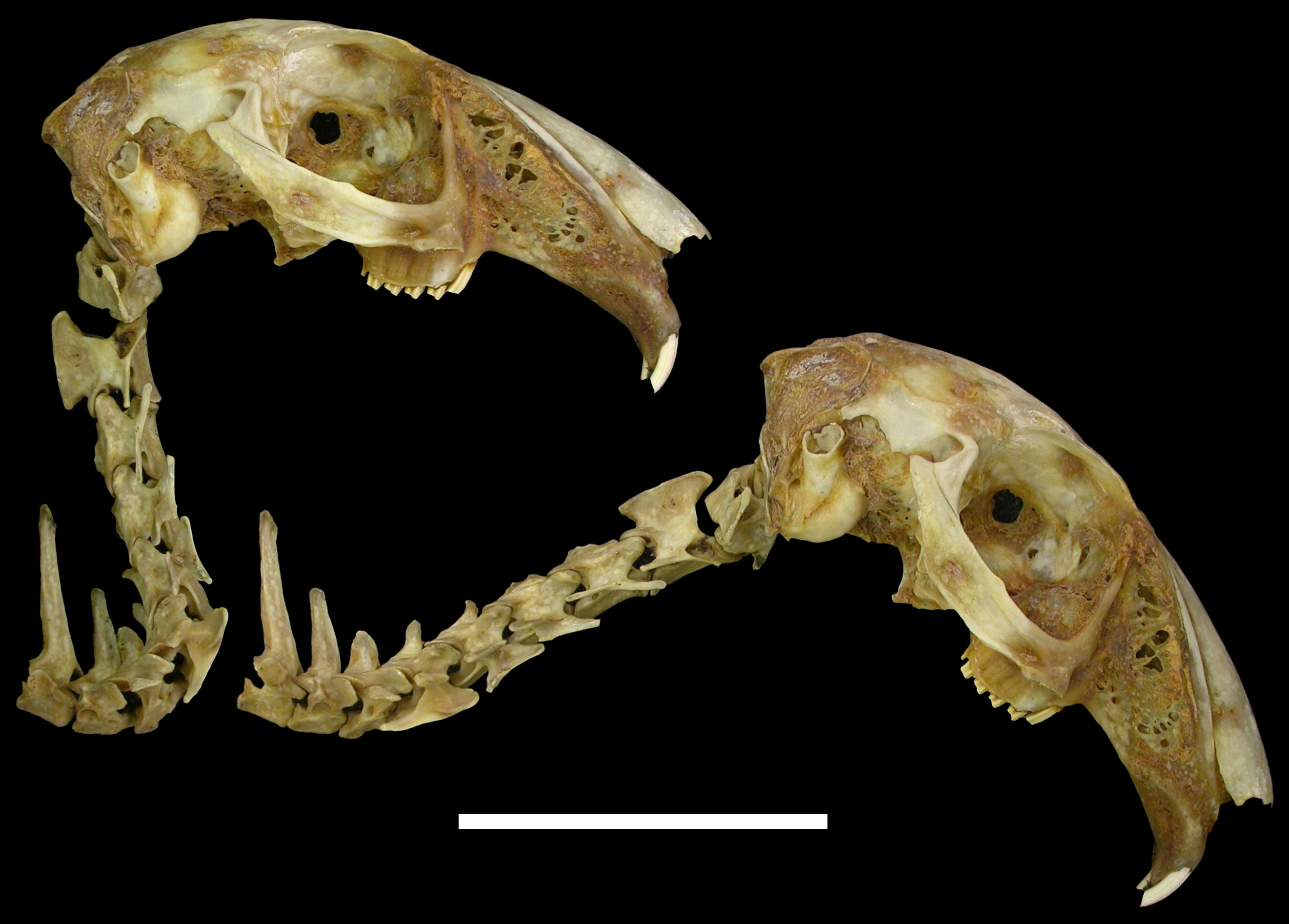
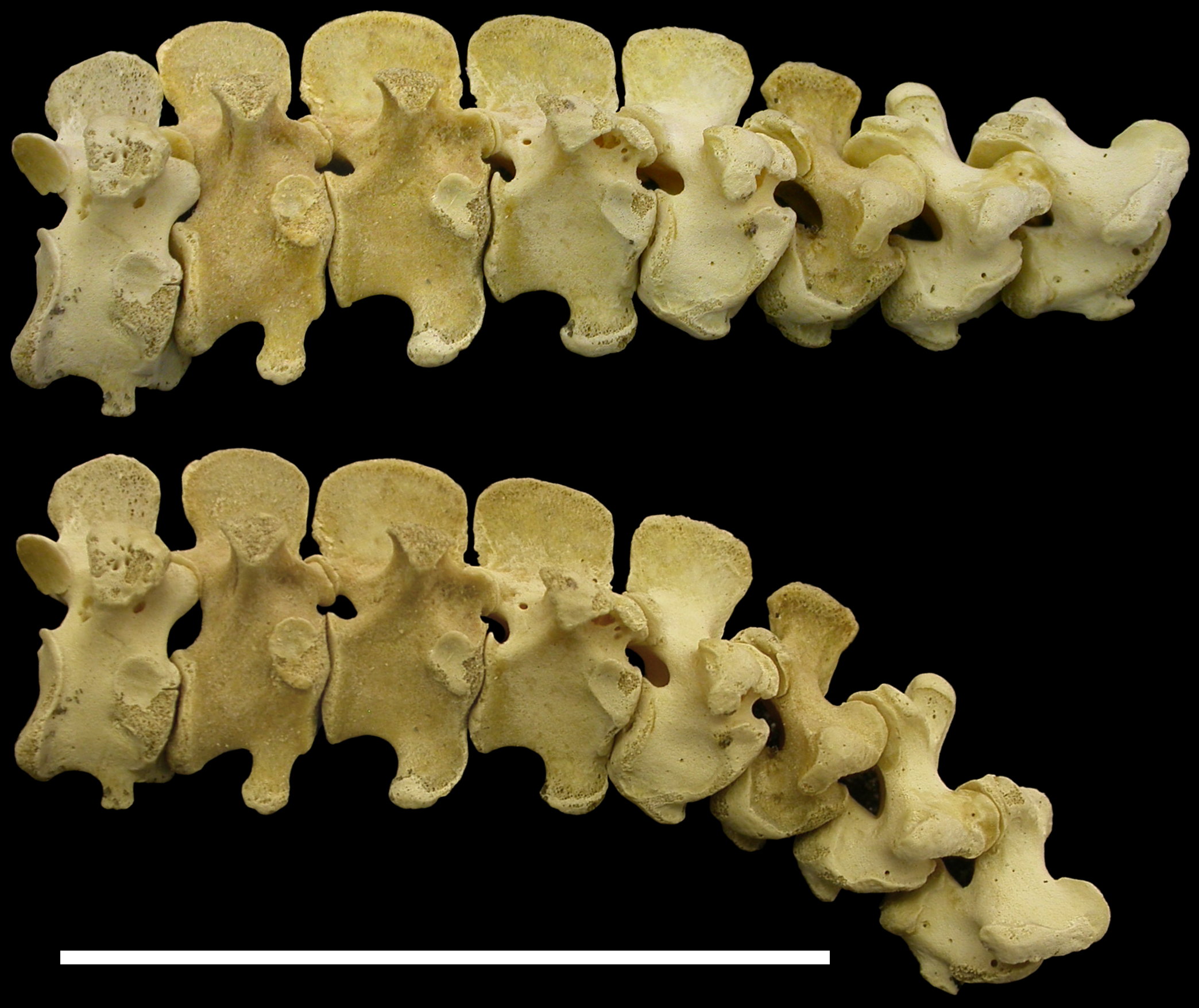
| Figure 1. Cape hare Lepus capensis RAM R2 in right lateral view, illustrating maximally extended pose and ONP: skull, cervical vertebrae 1-7 and dorsal vertebrae 1-2. Note the very weak dorsal deflection of the base of the neck in ONP, contrasting with the much stronger deflection illustrated in a live rabbit by Vidal et al. (1986: fig. 4). Scale bar 5 cm. |
|
| Figure 2. Chicken Gallus domesticus RAM R1 in right lateral view, illustrating maximally extended pose and ONP: last four cervical and first four dorsal vertebrae. Note the strong ventral deflection of the base of the neck in ONP, contrasting with the very strong dorsal deflection illustrated in a live chicken by Vidal et al. (1986: fig. 7). Scale bar 5 cm. |
|
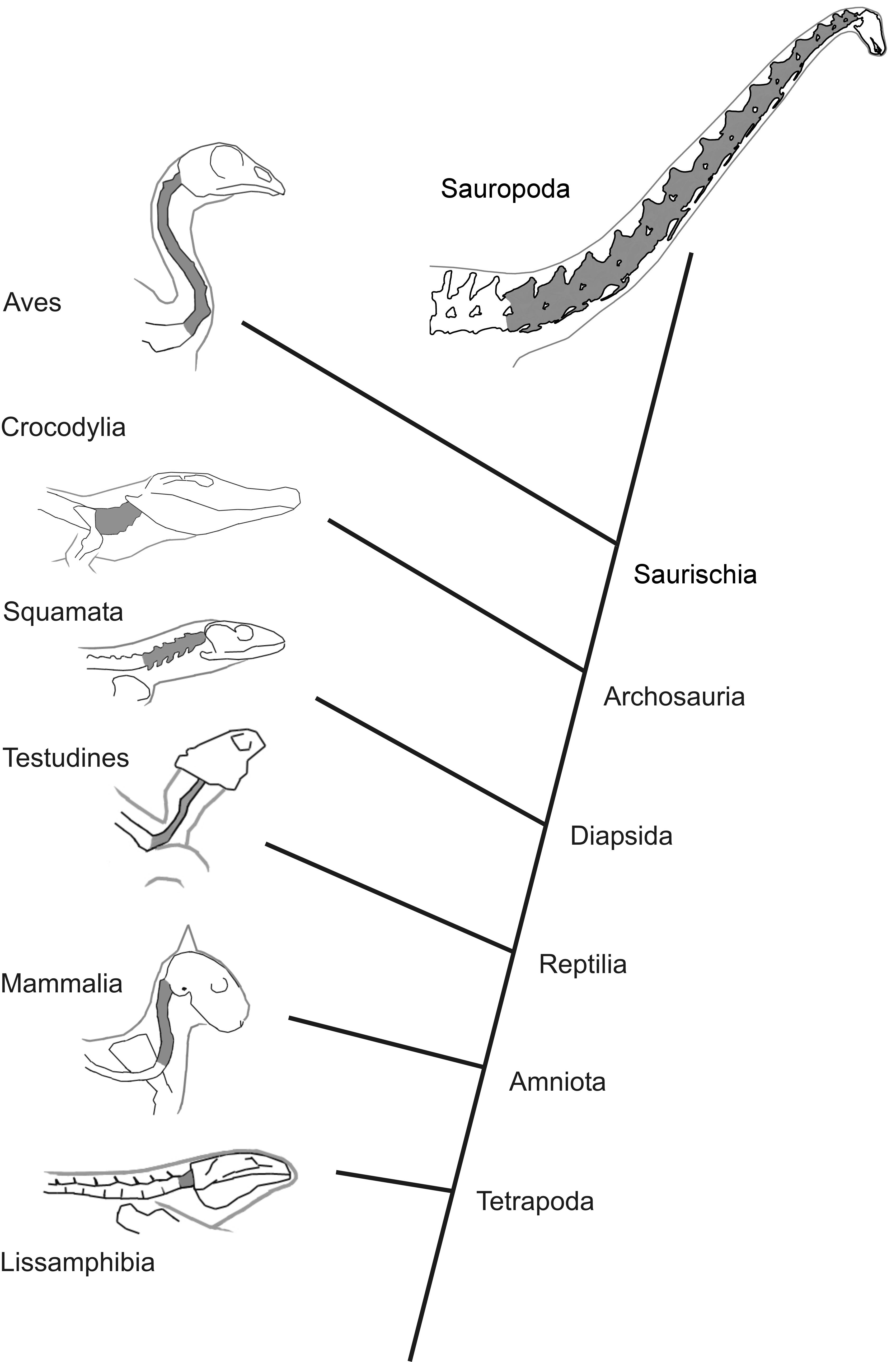
| Figure 3. Phylogeny indicating high-level relationships between tetrapod groups, habitual neck posture in extant groups, and inferred posture in sauropods. Cervical vertebrae shaded dark grey. Lissamphibia: Ambystoma tigrinum, after Simons et al. (2000: fig. 4); Mammalia: cat, after Vidal et al. (1986: fig. 3B); Testudines: Terrapene carolina, after Landberg et al. (2003, fig. 8); Squamata: Varanus exanthematicus, after Owerkowicz et al. (1999: fig. 2A); Crocodylia: alligator, after unpublished photograph; Aves: chicken, after Vidal et al. (1986: fig. 7); Sauropoda: Diplodocus carnegii, modelled after vertebrae in Hatcher (1901: fig. 4, pl. III). |
|
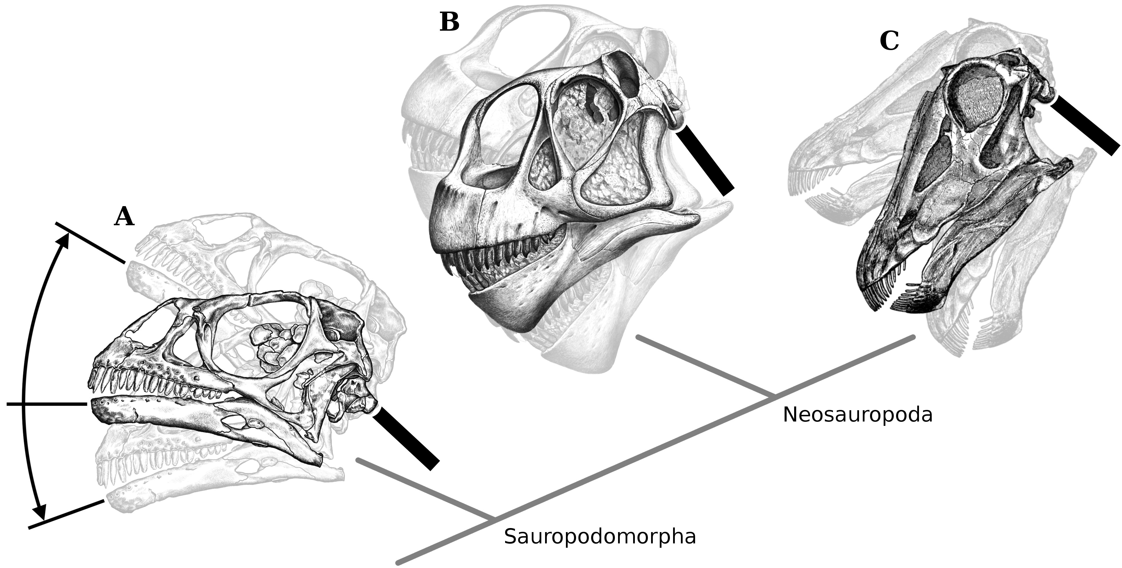
| Figure 4. Range of possible habitual head angles in the basal sauropodomorph A. Massospondylus, and the sauropods B. Camarasaurus and C. Diplodocus. Heads shown with HSSC oriented horizontally, and tilted 30º upwards and 20º downwards, the range of habitual orientations found for birds by Duijm (1951) . Necks shown in neutral position with respect to heads with horizontal HSCCs. Massospondylus BP/1/4376 after Sues et al. (2004: fig. 1A), Camarasaurus CM 11338 after Gilmore (1925: pl. XVI), Diplodocus USNM 2672 after Hatcher (1901: pl. II). |
|
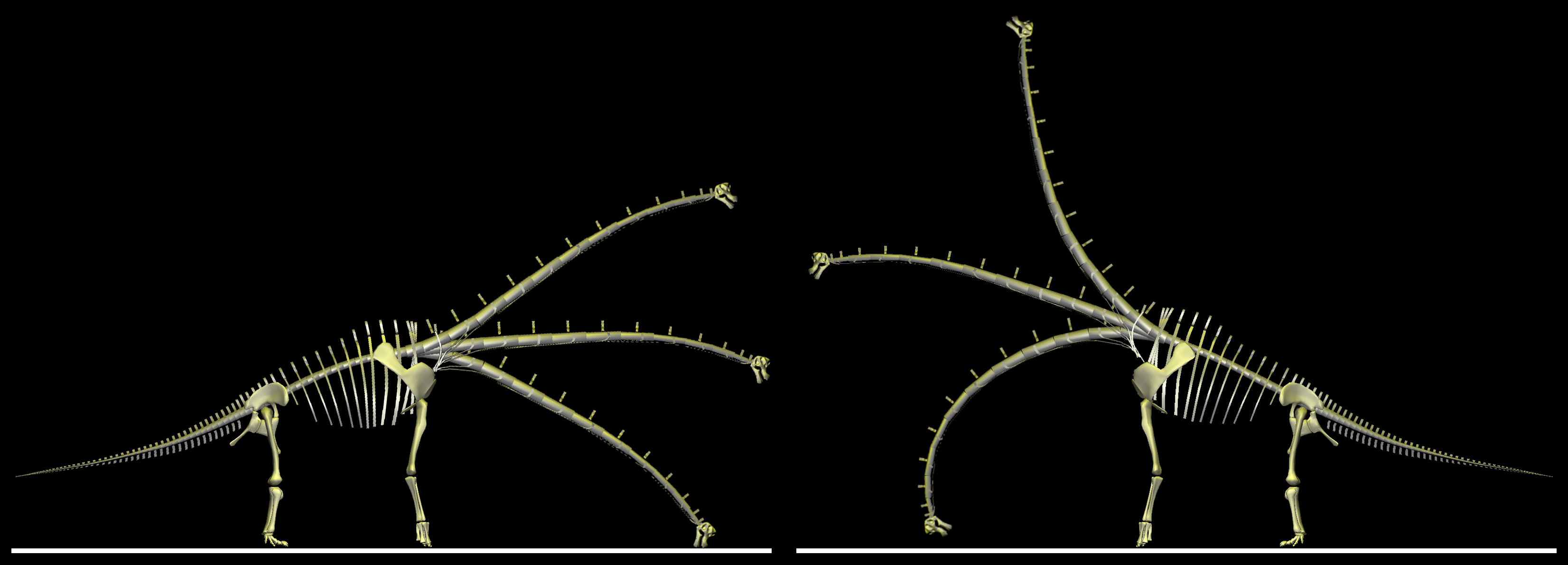
| Figure 5. Brachiosaurus brancai reconstructions with low and high torso positions. Neck in ONP, in a drinking posture, and in a browsing posture attained by deflecting the neck dorsally by the same amount as it is deflected ventrally to reach the ground. Torso, appendicular skeleton and ONP neck from Stevens and Parrish (2005b: fig. 6.8). Cervical joints deflected by 8º from ONP. See text for full details. |
|